
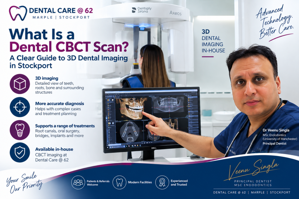
A conventional dental X-ray can show a great deal, but some questions need a clearer view. If you have a complex root canal problem, an impacted tooth, unexplained symptoms or are planning certain treatments, you may hear your dentist mention a CBCT scan. So, what is a dental CBCT scan? It is a type of dental X-ray that produces a detailed three-dimensional picture of your teeth, jawbone, roots, nerves and surrounding structures.
CBCT stands for cone beam computed tomography. Unlike a standard X-ray, which creates a flat image, a CBCT scanner takes many images as it moves around your head. Specialised software then combines them into a 3D image that the dentist can view from different angles and in fine detail.
For the right clinical situation, this additional information can support a more confident diagnosis and carefully planned treatment. It is not needed for every dental concern, and it is only recommended when the likely benefit justifies the extra radiation exposure.
At Dental Care @ 62 in Marple, Stockport, we have CBCT imaging available in-house. This means that where a 3D scan is clinically justified, it can form part of the assessment and treatment-planning process without the patient necessarily needing to travel elsewhere for imaging.
How does a dental CBCT scan differ from a normal X-ray?
Most routine dental X-rays are two-dimensional. Bitewing X-rays are particularly useful for checking for decay between teeth, while small periapical X-rays can show the root of an individual tooth and the bone around it. A panoramic X-ray, sometimes called an OPG, provides a broad overview of both jaws.
These images remain valuable and are often all that is required. However, a 2D image can sometimes leave uncertainty because structures overlap. For example, it may be difficult to tell whether a root is close to a nerve, whether a tooth has an extra root canal, or exactly how an unerupted tooth is positioned within the jaw.
A dental CBCT scan can address these questions by showing depth as well as height and width. The image can be examined in thin slices, rather like looking through the jaw layer by layer. This helps the clinician understand the relationship between teeth, bone, sinuses and important nerves before recommending the most appropriate next step.
When might a CBCT scan be recommended?
A CBCT scan is a diagnostic tool, not a routine add-on. Your dentist will first consider your symptoms, examination findings and any existing X-rays. If a standard image already answers the clinical question, a 3D scan may not be necessary.
There are circumstances where it can be particularly helpful, including:
- complex root canal assessment, especially where a tooth has unusual anatomy, persistent infection or previous treatment that has not settled as expected
- planning difficult extractions, including wisdom teeth that appear close to the nerve in the lower jaw
- assessing impacted or unerupted teeth before orthodontic treatment orthodontic treatment or surgical care
- planning dental implants by assessing the available bone and the location of nearby anatomical structures
- investigating certain jaw problems, fractures, cyst-like areas or unexplained changes seen on other X-rays
For someone with a failing root canal treatment, for instance, a CBCT image may show a hidden canal, a fracture pattern or the extent of infection around the roots more clearly than a conventional X-ray. That information does not automatically mean a tooth needs treatment or extraction. It helps the dentist discuss whether monitoring, further root canal treatment, surgery or another option may be suitable.
What happens during a dental CBCT scan?
Having a CBCT scan is straightforward and usually takes less than a minute to capture the images. There are no injections, enclosed tunnels or physical contact with the scanner.
You will usually be asked to remove glasses, jewellery, hearing aids or removable metal objects around the head and neck, as these can affect image quality. Depending on the equipment and area being scanned, you may sit or stand while the team carefully positions your head. A support helps you remain still as the scanner rotates around you.
The machine does not touch you. You may hear a quiet sound as it moves, but the scan itself is quick. The most useful thing you can do is keep still and follow the team’s instructions. If you are nervous, let the dental team know beforehand. They can explain each stage and make sure you are comfortable before starting.
Is a dental CBCT scan safe?
CBCT uses ionising radiation, as conventional dental X-rays do. This is why a scan should always have a clear clinical reason. Dental teams follow radiation protection guidance and use the smallest scan area and lowest exposure setting that is appropriate for the question being investigated.
The radiation dose varies considerably depending on the machine, scan size, settings and part of the mouth being examined. A small, focused scan usually involves less exposure than a large scan of both jaws. It is not helpful to compare every scan with a single everyday activity, because doses and circumstances vary. What matters is that the clinician weighs the potential diagnostic value against the exposure for you as an individual.
Tell your dentist if you are pregnant or think you might be pregnant. If dental imaging is clinically necessary, the dental team will consider the individual circumstances and ensure that the examination is appropriately justified. Children are also assessed with particular care, as they are more sensitive to radiation and should only have a CBCT scan when it is justified.
What can the scan show?
A CBCT scan is especially good at showing hard tissues, including teeth and bone. It can reveal the shape and number of tooth roots, fine details of root canal anatomy, bone levels, the position of impacted teeth and the proximity of roots to the lower jaw nerve or maxillary sinuses.
It can also help with treatment planning. Before an implant is considered, for example, the scan can help assess bone height and width. Before a difficult wisdom tooth removal, it can help identify whether the tooth roots appear close to the nerve that provides feeling to the lower lip and chin. Knowing this in advance allows a more informed conversation about the procedure, its alternatives and relevant risks.
There are limitations. CBCT is not generally the first choice for finding small areas of tooth decay, and it is less suited to assessing soft tissues than medical CT or MRI scans. Metal fillings, crowns and braces can create streaks or shadows that reduce clarity in parts of the image. A scan provides detailed information, but it still needs to be interpreted alongside your history, symptoms and clinical examination.
CBCT scans and dental referrals in Stockport
Dental Care @ 62 accepts referrals from other dentists for appropriate complex cases. Our in-house CBCT can support assessment and treatment planning where three-dimensional imaging is clinically justified, including selected endodontic, oral surgical and implant-related cases.
Dr Veenu Singla, Principal Dentist at Dental Care @ 62, holds an MSc in Endodontics from the University of Manchester and has a special interest in complex root canal treatment. CBCT imaging can be particularly valuable in selected endodontic cases where conventional X-rays do not provide enough information for diagnosis or treatment planning.
If you have been referred by your own dentist, the treating clinician will consider the referral information and explain whether a CBCT scan would add useful detail. The images are reviewed as part of your care, rather than being treated as a result in isolation. Where findings fall outside the immediate dental area, appropriate further advice or referral may be recommended.
Questions worth asking before a CBCT scan
Clear communication is part of good radiographic care. Your dentist should be able to explain why a 3D scan is being considered, what question it is intended to answer and whether there are alternatives. You may also wish to ask how the findings could affect your treatment options and whether the scan covers a small area or a wider section of the jaws.
For many patients, the reassurance comes from understanding that a CBCT scan is not being suggested simply because it is available. It is used when a more detailed view may change or improve the planning of care. Sometimes the scan confirms that a straightforward approach is appropriate. At other times, it identifies factors that need a more cautious plan.
Need a dental CBCT scan or complex dental assessment in Stockport?
Dental Care @ 62 in Marple has in-house CBCT imaging to support appropriate diagnosis and treatment planning for selected complex dental cases.
Whether you are a patient requiring further assessment or a dentist looking to refer a complex case, contact Dental Care @ 62 to discuss how we can help.
2 comments on “What Is a Dental CBCT Scan? 3D Dental Imaging in Stockport”
Options for Replacing Missing Teeth | Dental Care @ 62
[…] lost, especially if the gap has been present for some time. In appropriate cases, three-dimensional CBCT imaging can help assess the available bone and important nearby structures before treatment is planned. […]
Wisdom Tooth Removal Stockport | Dental Care @ 62
[…] chin, CBCT may provide additional three-dimensional information to help with treatment planning. CBCT is not routinely required before wisdom tooth removal and should only be used where clinically […]